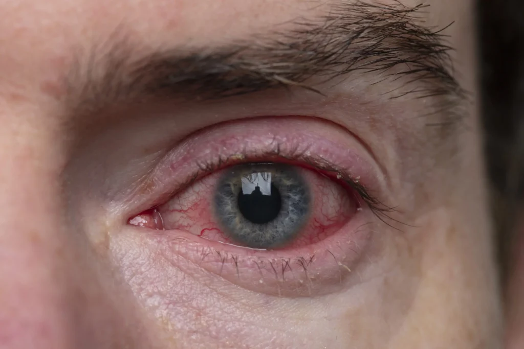
A corneal ulcer, also known as keratitis, is an open sore on the cornea—the transparent, dome-shaped layer covering the front of the eye. This condition can lead to severe pain, vision impairment, and potential blindness if left untreated. Corneal ulcers are commonly caused by infections but can also result from trauma, underlying health conditions, or prolonged contact lens use. Early detection and treatment are essential to prevent complications.

Causes of Corneal Ulcers
Corneal ulcers can arise due to infectious or non-infectious factors.
Infectious Causes
- Bacterial Infections
- Common bacteria involved include Pseudomonas aeruginosa, Staphylococcus aureus, and Streptococcus pneumoniae.
- Contact lens users are at higher risk due to bacterial contamination.
- Viral Infections
- The herpes simplex virus (HSV) can cause dendritic ulcers, leading to recurrent keratitis.
- The varicella-zoster virus, responsible for shingles, can also damage the cornea.
- Fungal Infections
- Fungal keratitis is caused by fungi such as Fusarium, Aspergillus, and Candida.
- It often follows eye trauma involving plant material or occurs in immunocompromised individuals.
- Parasitic Infections
- Acanthamoeba keratitis is a severe infection primarily affecting contact lens wearers exposed to contaminated water.
Non-Infectious Causes
- Dry Eye Syndrome – Severe dryness can lead to corneal surface damage.
- Physical Trauma – Injury from foreign bodies, chemical burns, or surgery can trigger ulcer formation.
- Autoimmune Diseases – Conditions such as rheumatoid arthritis can cause peripheral ulcerative keratitis, leading to corneal thinning.
Symptoms of Corneal Ulcers
Patients with corneal ulcers may experience:
- Severe eye pain and discomfort
- Redness and inflammation
- Excessive tearing
- Eye discharge (pus or mucus)
- Blurred or reduced vision
- Increased sensitivity to light (photophobia)
- A visible white or gray spot on the cornea
- Swelling of the eyelids
In advanced cases, the ulcer may deepen, leading to corneal perforation, a medical emergency that requires immediate intervention.
Diagnosis of Corneal Ulcers
A thorough eye examination is essential for accurate diagnosis.
- Slit-Lamp Examination – A specialized microscope is used to examine the cornea for ulcer size, depth, and location.
- Fluorescein Staining – A special dye highlights corneal defects, allowing better visualization of ulcers.
- Microbiological Cultures – Scraping the ulcer for laboratory analysis helps identify bacterial, fungal, or viral pathogens.
- Corneal Imaging – Optical coherence tomography (OCT) may be used to assess deeper corneal involvement.
Treatment of Corneal Ulcers
Medical Treatment
- Antibiotic Eye Drops – Broad-spectrum antibiotics are prescribed for bacterial infections.
- Antiviral Medications – Antiviral drops or oral medications help treat herpes-related ulcers.
- Antifungal Therapy – Topical antifungal agents are used for fungal keratitis.
- Pain Management – Cycloplegic eye drops help relieve discomfort and photophobia.
Surgical Interventions
For severe or unresponsive cases, surgical procedures may be required:
- Debridement – Removal of infected or necrotic tissue to promote healing.
- Conjunctival Flap Surgery – Covering the ulcer with conjunctival tissue to aid in recovery.
- Corneal Transplantation (Keratoplasty) – In extreme cases, a damaged cornea may need replacement with donor tissue.
Prevention of Corneal Ulcers
Preventative measures can significantly reduce the risk of corneal ulcers:
- Proper Contact Lens Hygiene – Clean and store lenses as recommended; avoid sleeping in contact lenses.
- Eye Protection – Use safety goggles during activities that may cause eye injury.
- Prompt Treatment of Eye Conditions – Seek medical attention for eye infections, injuries, or persistent dryness.
- Regular Eye Checkups – Routine examinations help detect early signs of corneal damage.

