Cytomegalovirus (CMV) retinitis is the most common opportunistic ocular infection in patients with Acquired Immunodeficiency Syndrome (AIDS). It primarily occurs in individuals with severe immunosuppression, particularly those with a CD4+ T-cell count below 50 cells/µL. Left untreated, CMV retinitis can lead to blindness due to retinal necrosis and detachment. This article explores the epidemiology, clinical presentation, diagnosis, and treatment of CMV retinitis in AIDS patients.

Epidemiology of CMV Retinitis in AIDS Patients
Before the introduction of highly active antiretroviral therapy (HAART), CMV retinitis affected nearly 30% of AIDS patients. With the advent of HAART, its incidence has declined significantly, though it remains a major cause of vision loss in advanced HIV cases.
Risk Factors
- Severe Immunosuppression: CD4+ count below 50 cells/µL
- HAART Non-Adherence: Patients not on effective antiretroviral therapy
- Previous CMV Infection: Reactivation of latent CMV infection
- Advanced HIV Disease: Late-stage AIDS with multiple opportunistic infections
Clinical Manifestations of CMV Retinitis
CMV retinitis typically presents unilaterally but can progress to bilateral involvement if untreated. Symptoms develop gradually and may include:
Common Symptoms
- Floaters: Dark spots moving across the visual field
- Blurred Vision: Progressive loss of clarity
- Photopsias: Sensation of flashes of light
- Visual Field Loss: Peripheral or central vision defects
Ophthalmic Examination Findings
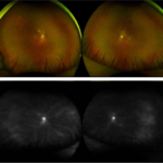
- Yellow-White Retinal Lesions: Areas of necrosis with granular borders
- Perivascular Hemorrhages: Often referred to as the “pizza pie” or “cheese and ketchup” appearance
- Mild Vitritis: Minimal inflammation in the vitreous compared to other infectious retinitis
Diagnosis of cmv retinitis in aids patient
Clinical Diagnosis
The diagnosis is primarily clinical, based on a dilated fundoscopic examination. Ophthalmologists identify the characteristic retinal changes associated with CMV retinitis.
Laboratory and Imaging Tests
- Polymerase Chain Reaction (PCR): Detects CMV DNA in blood or ocular fluids
- Fluorescein Angiography: Identifies areas of ischemia and vascular leakage
- Optical Coherence Tomography (OCT): Evaluates retinal thinning and atrophy
Treatment of CMV Retinitis
Timely intervention is critical to prevent irreversible vision loss. Treatment involves systemic and/or intravitreal antiviral therapy.
Antiviral Medications
| Drug | Route | Side Effects |
|---|---|---|
| Ganciclovir | Intravenous, Intravitreal | Bone marrow suppression, nephrotoxicity |
| Valganciclovir | Oral | Nausea, diarrhea, neutropenia |
| Foscarnet | Intravenous, Intravitreal | Nephrotoxicity, electrolyte imbalance |
| Cidofovir | Intravenous | Nephrotoxicity, uveitis, hypotony |
HAART and Immune Recovery
- Effective HAART is essential in restoring immune function and reducing CMV recurrence.
- Patients with a sustained CD4+ count above 100 cells/µL for at least three months may discontinue CMV maintenance therapy.
Prevention Strategies
- Regular Ophthalmic Screening: Early detection in at-risk individuals
- Strict HAART Adherence: Maintains immune function and prevents CMV reactivation
- Prophylactic Therapy: Considered for high-risk patients with extremely low CD4+ counts
CMV retinitis remains a serious opportunistic infection in AIDS patients with profound immunosuppression. While HAART has significantly reduced its incidence, timely diagnosis and appropriate antiviral therapy are crucial in preserving vision. Regular ophthalmologic monitoring and strict adherence to antiretroviral therapy are the most effective strategies for preventing CMV-related blindness.